Team Ducktectors
Oncoscope
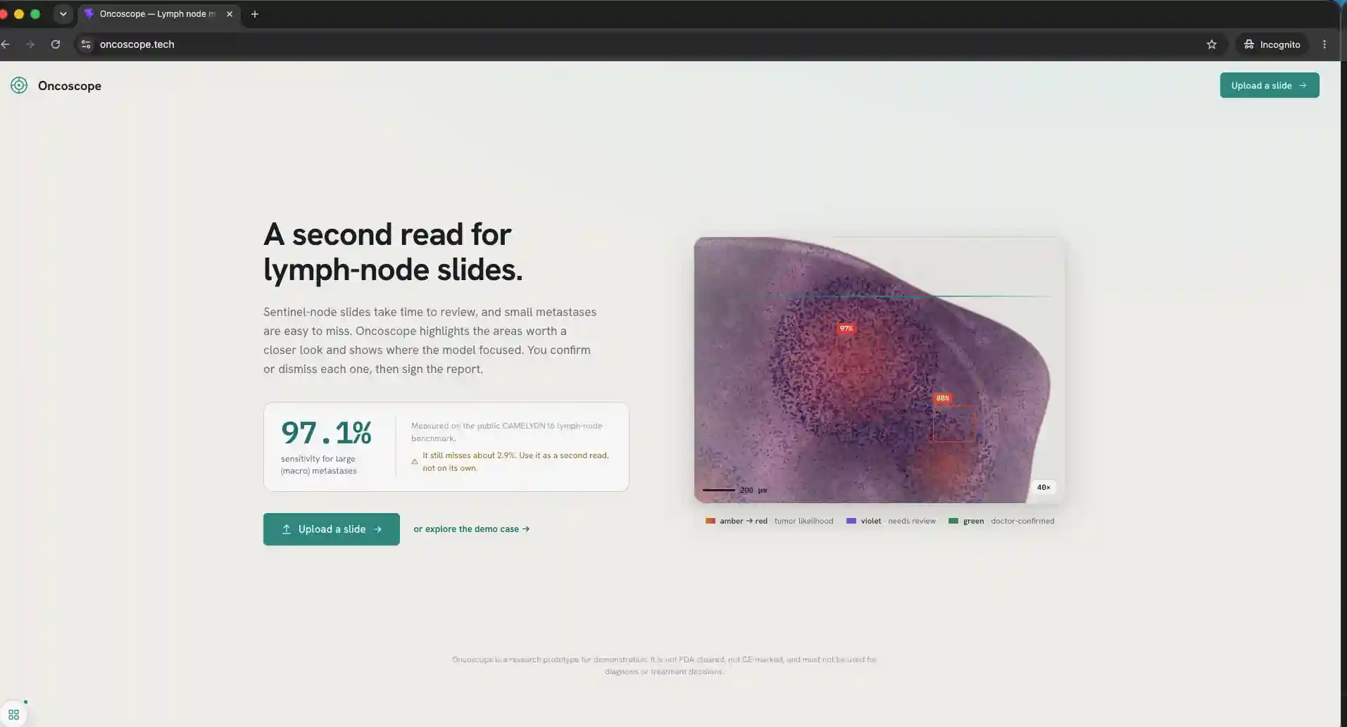
A Copilot for Pathologist
About this project
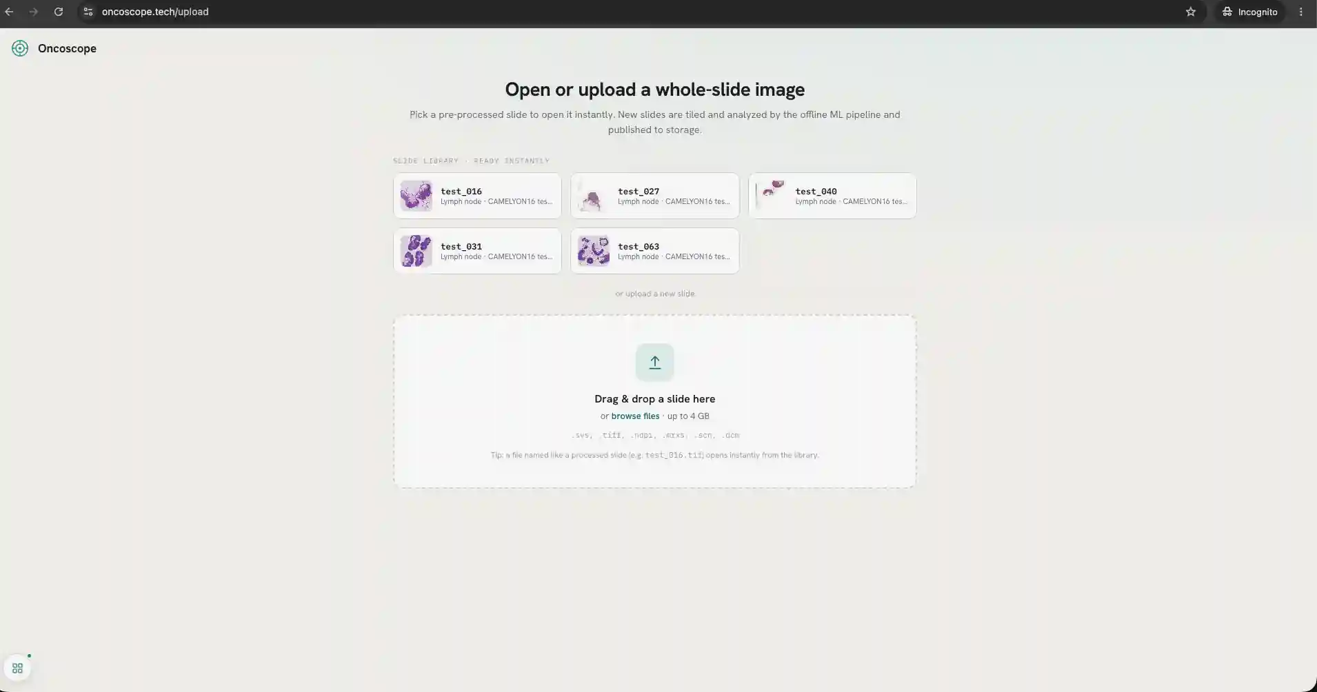
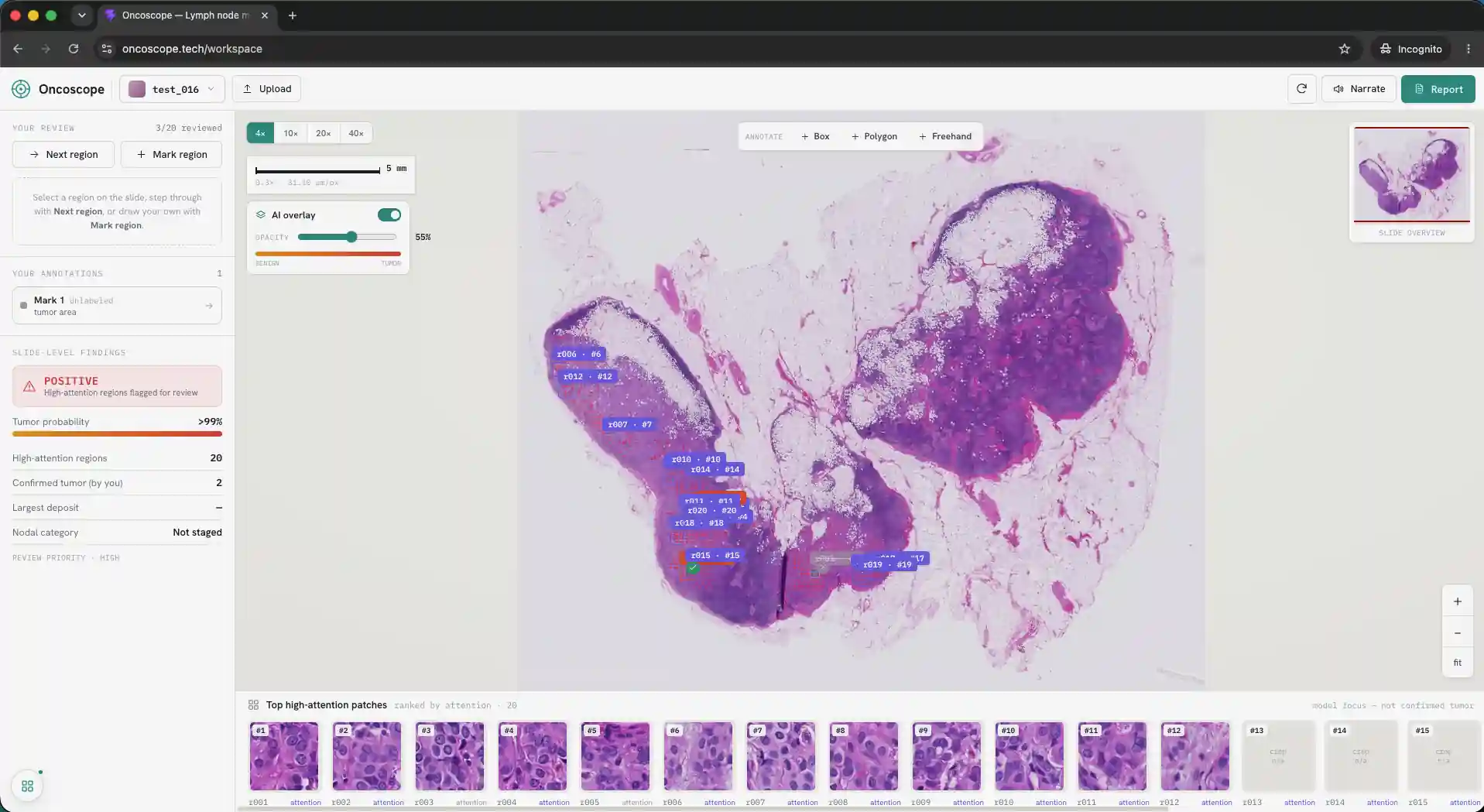
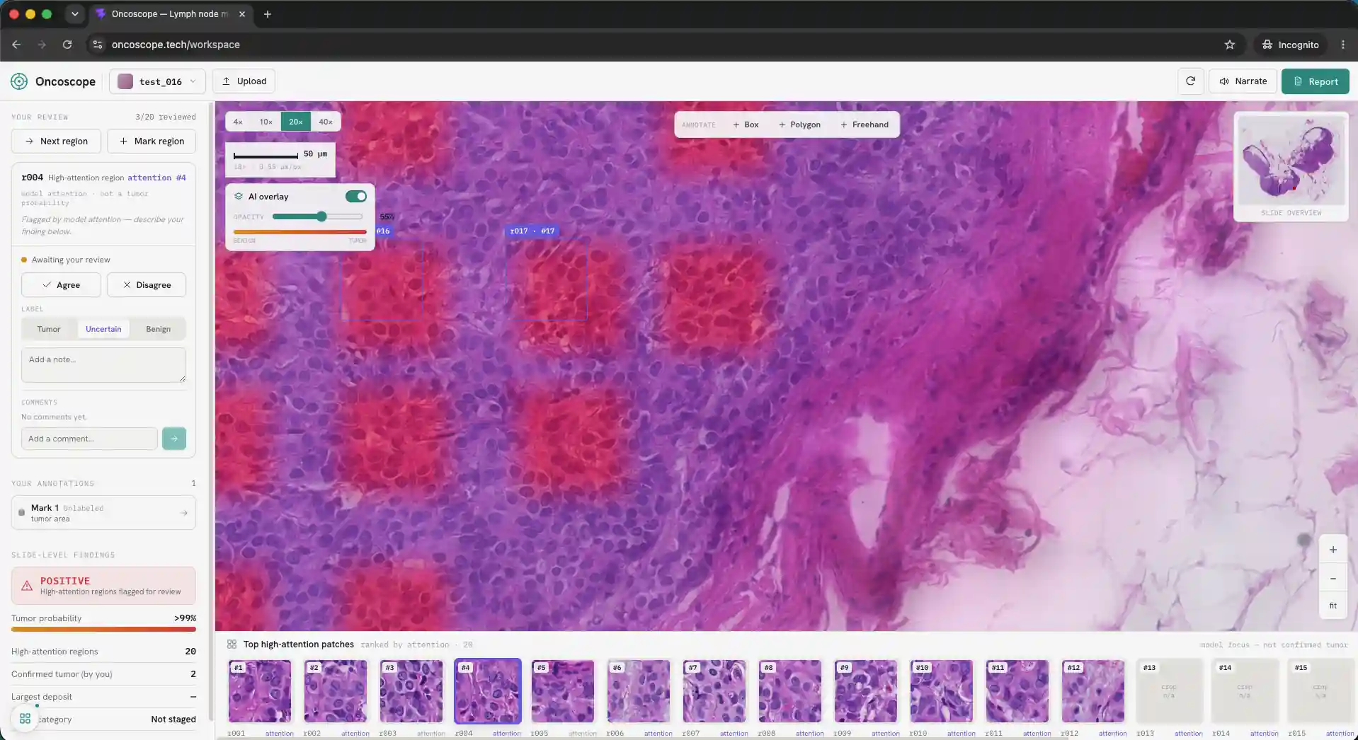
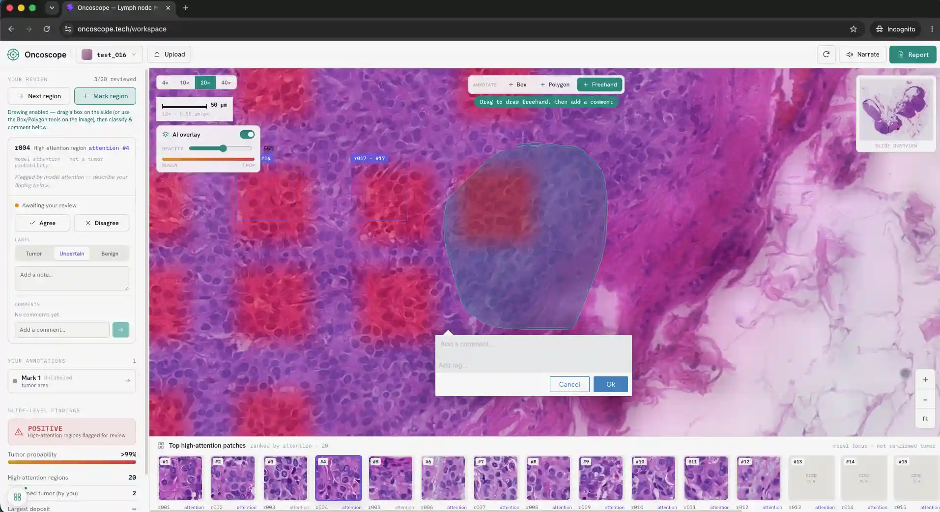
We have a project on cancer detection in whole slide images. This is not simple cancer classification; it is tumor detection and localization at very high resolution. Each whole slide image can be around 1 GB, so even loading the image in a browser is difficult. We built the system so pathologists can smoothly view, zoom, review, and verify AI-highlighted regions. We have three main goals: - Reduce missed cancers. - Make the AI’s evidence visible and verifiable. - Keep the pathologist in control. According to People Beating Cancer, roughly 11% of cancer patients are misdiagnosed, meaning for every 1,000 people, around 110 may experience a cancer misdiagnosis in their lifetime. Our goal is not to replace the pathologist, but to support them by highlighting suspicious regions that may otherwise be missed during review. Our main target is to reduce false negative by making every AI result reviewable, editable, and explainable. ## Training a model Each hospital can have different staining protocols, scanners, tissue preparation methods, and image quality. - We used a large foundation model because it already understands many visual patterns. - Instead of training a full model from scratch, we trained a classifier on top of foundation-model embeddings. - This lets us adapt the system to tumor detection while keeping training fast. - We used an NVIDIA H200 GPU and optimized the workflow to finish training in around 10 minutes. - The long-term idea is that this can be customized for different cancer types and different hospital data. ## Building the viewer ### Creating Tiles Initially, each WSI was around 1.1 GB, which cannot be loaded directly in the browser. - We converted the WSI into Deep Zoom Image tiles. - The slide is split into many small image tiles at different zoom levels. - The browser only loads the tiles needed for the current view. - This makes zooming and panning smooth, even for very large slides. - We used Goo
Gallery